Analytical Services Core
About
In 2021, the Histology, Microscopy and Imaging Core was transformed into the Analytical Services (AS) Core. The Analytical Services Core provides the resources and expertise for histology, microscopy, and mass spectrometry services as well as access to the tissue culture equipment.
The AS Core has three managers: Ms. Catherine Doller and Ms. Dawn Smith who provide histology services, and Ms. Maryanne Pendergast who provides training and access to high end microscopes, imaging software, and the expertise needed to successfully create microscopy-based studies. In addition, Ms. Smith oversees the use of tissue culture and other equipment, which is available through the Core. Access to mass spectrometry services is offered through a collaboration with Dr. Marcin Golczak, a faculty member in Pharmacology.
Histology
The Histology component of the Core does embedding and sectioning of tissue samples (both frozen and paraffin), usually of very precise areas (for example the optic nerve). During consultation, the Core Managers will advise whether to either fix tissues and embed (frozen) or to process and embed in paraffin. The Histology component also offers histological stains of the resulting slides. Routine H&E stain is available, as well as more complex stains such as Masson’s trichrome and Periodic Acid/Schiff’s.
How to Utilize:
-
Please contact either Cathy Doller at 216.368.5239 catherine.doller@case.edu, or Dawn Smith at 216.368.0790 dawn.smith@case.edu prior to beginning projects.
-
Complete the Histology Work Request Form.
For pricing information please contact either Cathy Doller at 216.368.5239 catherine.doller@case.edu or Dawn Smith at 216.368.0790 dawn.smith@case.edu.
Microscopy and Imaging
The Microscopy and Imaging component of the Analytical Services Core provides access and training in the correct use of the four microscope systems housed in the facility:
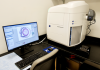
- The Zeiss Axio Scan.Z1 Slide Scanner facilitates the rapid and automated digitization of both bright-field and fluorescent slides. The ZEN Blue software supports the creation of course and fine focus maps, the acquisition and stitching of tiles, pyramidal viewing,
acquisition of z-stacks and a myriad of post processing options including the creation of Extended Depth of Focus images from the z-stacks.
-
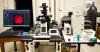
The Olympus FV1200 Confocal Microscope is based on an inverted platform and has five lasers (405nm, 440nm, 473nm, 559nm, 635nm) that can excite most conventional fluorescent dyes. The user-friendly FluoView software supports the acquisition of z-stacks, tiled images and time-lapse experiments in standard size dishes and multi-well plates.
-
The Leica DMI 6000b Inverted Microscope is an automated wide-field system with a temperature-controlled CO2 incubator that supports extended time-lapse experiments and the acquisition of BF, phase, and fluorescent images. In addition to the standard R, G, and B, fluorescent filter sets, the scope also has CFP/YFP cubes that support FRET experiments. The acquisition software is Metamorph.
-
The Olympus BX-60 Upright Microscope is a manual wide-field system with BF, fluorescence, and DIC capabilities that can acquire both grey scale and color images. The acquisition software is Metamorph and the module’s deconvolution software, AutoQuant, is accessed through this scope's version of the application.

Users can also be instructed in the proper use of any scope they may have in their own labs.
Metamorph is a very flexible and powerful image analysis application. The creation of automated macros (journals) within Metamorph not only speeds up the pace of image acquisition but also provides consistent results when analyzing large data sets. In addition to the two acquisition copies of Metamorph, the module also has five offline licenses that allow remote access to Metamorph on the offline server. The software can be loaded onto any Win10 PC and then accessed at the user’s convenience at home or in the lab. Image analysis services and training is also provided.
Users are strongly encouraged to contact the module’s manager prior to beginning any microscopy-based experiments. A review of experimental design, the correct selection of dyes and which microscope will best do the job are all-important factors to consider.
For additional or pricing information please contact Maryanne Pendergast at 216.368.4821 or mxp19@case.edu.
Tissue Culture Equipment
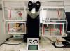
The Core provides access to the following equipment:
- Three biological safety cabinets Class II, Type A
- Six Thermo Scientific CO2 incubators
- An Amsco Lab 110 Sterilizer
- Two Leica DMil inverted microscopes
- A Nikon TS100 inverted microscope with fluorescence and phase contrast
- A Leica S4E Stereo microscope
- Nikon Eclipse 200 Microscope
- An Eppendorf 5810R refrigerated centrifuge
- An Eppendorf 5804 tabletop centrifuge
- Three Thermolyne 4 Plus cryobiological storage vessels
- An Amaxa Nucleofector II
- A Bio-Rad TC20 Automated Cell Counter
- An AirClean 600 PCR Workstation
- MilliQ water system (with Elix deionizer and a Synthesis A10 reverse osmosis system)
To utilize or for pricing information please contact Dawn Smith at 216.368.0790 or dawn.smith@case.edu
Mass Spectrometry
The Mass Spectrometry facility has the capability to provide the following services:
• identification and quantification of small-molecule metabolites in ocular tissues
• assessment of lipid profiles in mouse models of human conditions
• determination of pharmacokinetics and tissue distribution of experimental drugs
• determination of catalytic activity and substrate specificity of enzymes involved in visual processes
• determination of catalytic mechanism of enzymatic reactions by heavy atom labeling
• assessment of protein dynamics by hydrogen/deuterium exchange methodology
• analysis of binding specificity of lipid-transporting proteins
To utilize please contact Dr. Marcin Golczak at 216.368.0302 or marcin.golczak@case.edu
Contact Information
Histology Office
Catherine Doller
Institute of Pathology Building, Room 108
Email: catherine.doller@case.edu
Phone: 216.368.5239
Dawn Smith
Institute of Pathology Building, Room 108
Email: dawn.smith@case.edu
Phone: 216.368.0790
Microscopy and Imaging Office
Maryanne Pendergast
Institute of Pathology Building, Room 105
Email: mxp19@case.edu
Phone: 216.368.4821
Tissue Culture Equipment Office
Dawn Smith
Institute of Pathology Building, Room 108
Email: dawn.smith@case.edu
Phone: 216.368.0790
Mass Spectrometry Office
Marcin Golczak, Ph.D.
Department of Pharmacology
Wood Building, Room W151C
Email: marcin.golczak@case.edu
Phone: 216.368.0302
Module Co-directors
Carlos Subauste, M.D.
Departments of Medicine and Pathology
Biomedical Research Building 448A
Email: carlos.subauste@case.edu
Phone: 216.368.2785
Patricia Taylor, Ph.D.
Department of Ophthalmology and Visual Sciences
Pathology 101
Email: patricia.r.taylor@case.edu
Phone: 216.368.3171