Research
Nanomaterials to Improve Energy Conversion Schemes
Nanoscience leads to a wide range of energy applications from photovoltaics to thermoelectrics. It is expected that nanostructures play a pivotal role in the quickly rising areas of energy conversion and energy storage. We synthesize inorganic nanocrystals and control their sizes, compositions, and their surfaces and characterize them with state-of-the-art technology, including electron microscopy with a 1 nanometer resolution and optical spectroscopy with a time-resolution of 100 femotseconds to uncover the often surprising properties of the size-confined materials. For example, we investigate the optoelectronic properties of nanocrystals and develop nanomaterials with properties of interest for the complex nanoarchitectures needed in energy-related applications. Please consult our publications for an update on recent research.
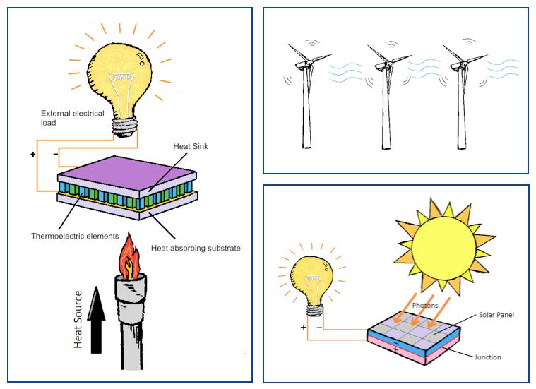
Image credit: Michael Chiang
References:
- Sang, Lixia; Tan, Huanyue; Zhang, Xiaomin; Wu, Yuting; Ma, Chongfang; Burda, Clemens. "Effect of Quantum Dot Deposition on the Interfacial Flatband Potential, Depletion Layer in TiO2 Nanotube Electrodes, and Resulting H2 Generation Rates". Journal of Physical Chemistry C (2012), 116(35), 18633-18640.
- Zhao, Yixin; Burda, Clemens. "Development of plasmonic semiconductor nanomaterials with copper chalcogenides for a future with sustainable energy materials". Energy & Environmental Science (2012).
- Zhao, Yixin; Dyck, Jeffrey S.; Hernandez, Brett M.; Burda, Clemens. "Improving Thermoelectric Properties of Chemically Synthesized Bi2Te3-Based Nanocrystals by Annealing". Journal of Physical Chemistry C (2010), 114(26), 11607-11613.
Optoelectronics of Organic and Inorganic Material
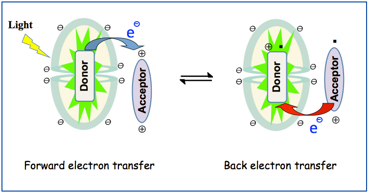
Organic materials are the basis of increasingly many materials and devices. However the electronic properties of organic materials, though fascinating, are complex and require detailed spectroscopic investigation. With femtosecond time-resolved laser spectroscopy and imaging techniques, studies of ensembles of molecules are performed. The goal of this research is to increase the understanding of the chemical dynamics and the electronic properties of molecules, assemblies, and nanocomposites. The uncovered novel concepts will be guide for the design of new technological or biomedical prototype devices. We are particularly interested in understanding the interfacing between molecules, nano-structures, and macroscopic materials. Building functional devices from nanostructures requires an understanding of the interactions between these components.
References:
- Porel, Mintu; Chuang, Chi-Hung; Burda, Clemens; Ramamurthy, Vaidhyanathan. "Ultrafast Photoinduced Electron Transfer between an Incarcerated Donor and a Free Acceptor in Aqueous Solution". Journal of the American Chemical Society (2012), 134(36), 14718-14721.
- Lou, Yongbing; Chen, Xiaobo; Samia, Anna C.; Burda, Clemens. "Femtosecond Spectroscopic Investigation of the Carrier Lifetimes in Digenite Quantum Dots and Discrimination of the Electron and Hole Dynamics via Ultrafast Interfacial Electron Transfer". Journal of Physical Chemistry B (2003), 107(45), 12431-12437.
- Mao, Baodong; Chuang, Chi-Hung; Lu, Feng; Sang, Lixia; Zhu, Junjie; Burda, Clemens. "Study of the Partial Ag-to-Zn Cation Exchange in AgInS2/ZnS Nanocrystals". Journal of Physical Chemistry C (2013), 117(1), 648-656.
Environmental Studies
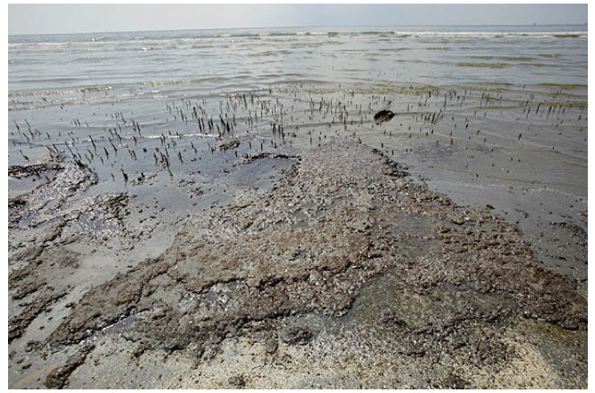
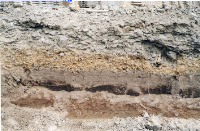
Persistent organic pollutants are a pressing issue for our environment. These compounds are resistant to environmental degradation through chemical, biological, and photolytic processes. They have been observed to persist in the environment, are capable of long-range transport, and exhibit bioaccumulation in human and animal tissue as well as in food chains. Therefore, they have potentially a significant impact on human health and the environment. We study the possibility to photodegrade organic pollutants in aqueous and lipophilic environments using the help of metal oxide nanostructures (TiO2, CeO2, etc.). We synthesize these photocatalysts, we dope them to achieve visible-light activation (e.g. using sun light to clean up oil spills, to clean surfaces under ambient light, etc.) and study the photoconversion of the pollutants into their photooxidation products. Often these decompositions are completely mineralizing the pollutants into CO2 and other inorganic oxidation products.
 |
 |
 |
References:
- Zhao, Yixin; Burda, Clemens. "Development of plasmonic semiconductor nanomaterials with copper chalcogenides for a future with sustainable energy materials". Energy & Environmental Science (2012), 5(2), 5564-5576.
- Doane, Tennyson L.; Cheng, Yu; Babar, Amir; Hill, Reghan J.; Burda, Clemens. "Electrophoretic Mobilities of PEGylated Gold NPs". Journal of the American Chemical Society (2010), 132(44), 15624-15631.
- Liu, Y.; Li, J.; Qiu, X.; Burda, C. "Novel TiO2 nanocatalysts for wastewater purification: tapping energy from the sun". Water Science and Technology (2006), 54(8, 5th World Water Congress: Wastewater Treatment Processes, 2006).
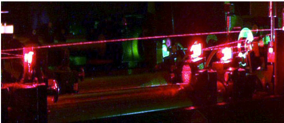
Ultrafast Laser Spectroscopy & Imaging
The latest laser and imaging technology is available in our Center for Chemical Dynamics and additional facilities at CWRU to reveal the electronic and morphological properties of nanostructures in materials, cells, and prototype device structures. State-of-the-art femtosecond laser spectroscopy and imaging are used to uncover the relevant photoinduced processes. This is a promising field that aides in developing new and ambitious ideas in the context of energy, environment, and health-related research.
References:
- Chi-Hung; Chen, Xiaobo; Burda, Clemens. "Femtosecond time-resolved hot carrier energy distributions of photoexcited semiconductor quantum dots". Annalen der Physik (Berlin, Germany) (2013), 525(1-2), 43-48.
- Doane, Tennyson L.; Chuang, Chi-Hung; Chomas, Andrew; Burda, Clemens, "Photophysics of Silicon Phthalocyanines in Aqueous Media". ChemPhysChem (2013), 14(2), 321-330.
- Chuang, Chi-Hung; Burda, Clemens. "Contribution of Femtosecond Laser Spectroscopy to the Development of Advanced Optoelectronic Nanomaterials". Journal of Physical Chemistry Letters (2012), 3(14), 1921-1927.
Nanoparticles for Health Applications
We are undertaking a critical assessment of the use of quantum dots and metallic nanoparticles in the delivery of drugs and therapy. A major advantage of using QDs is their strong photoluminescence, which in principle can be exploited for imaging and photodynamic therapy applications. This also entails modifying these nanostructures in their size, shape and surface composition. Studies are being carried out in cells and in vivo at the University Hospitals on campus.
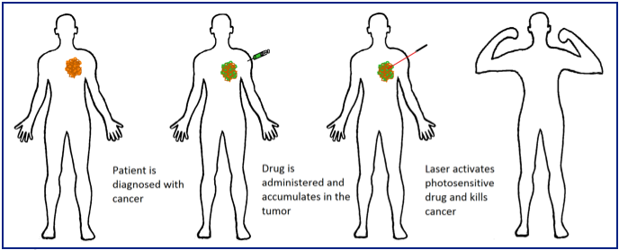
Image credit: Michael Chiang
References:
- Cheng, Yu; Meyers, Joseph D.; Agnes, Richard S.; Doane, Tennyson L.; Kenney, Malcolm E.; Broome, Ann-Marie; Burda, Clemens; Basilion, James P. "Addressing Brain Tumors with Targeted Gold Nanoparticles: A New Gold Standard for Hydrophobic Drug Delivery?". Small (2011), 7(16), 2301-2306.
- Doane, Tennyson L.; Burda, Clemens. "The unique role of nanoparticles in nanomedicine: imaging, drug delivery and therapy". Chemical Society Reviews (2012), 41(7), 2885-2911.
- Cheng, Yu; Meyers, Joseph D.; Broome, Ann-Marie; Kenney, Malcolm E.; Basilion, James P.; Burda, Clemens. "Deep Penetration of a PDT Drug into Tumors by Noncovalent Drug-Gold Nanoparticle Conjugates". Journal of the American Chemical Society (2011), 133(8), 2583-2591.