

Education
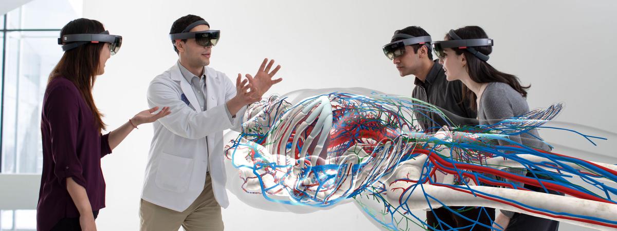
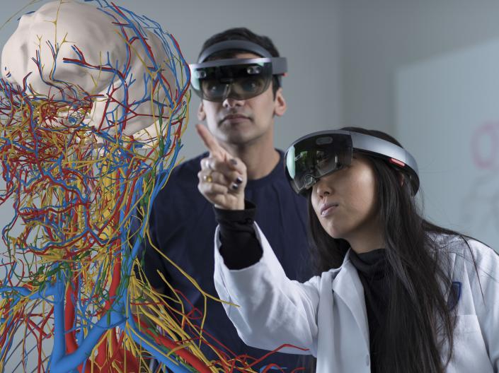
Department of Anatomy faculty are leaders in the creation of new ways of training tomorrow’s doctors, dentists, and physician assistants including the use of augmented reality in learning human anatomy.

MS in Applied Anatomy
In the Master of Science in Applied Anatomy program, our students broaden and deepen their knowledge of basic biology, human health and disease while gaining hands-on experiences to better prepare themselves for a biomedical career.
Research
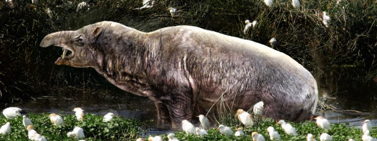
Faculty are advancing our knowledge of the evolution of mammals and humans, human anatomical variation and functional anatomy, and in medical education.
Join us for an information session
Sign up for upcoming events below!
Want to learn more?
Get to know our MS in Applied Anatomy
Join our Prestigious Faculty
The Anatomy Department has an opening for an Assistant/Associate Professor in Neuroanatomy
AAMC Premed Navigator
